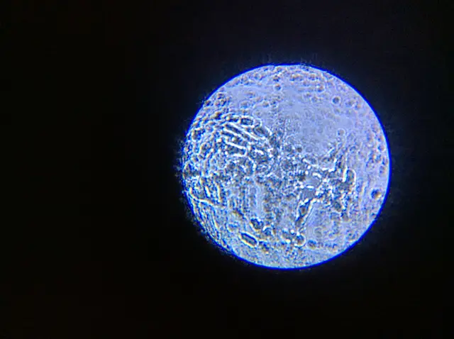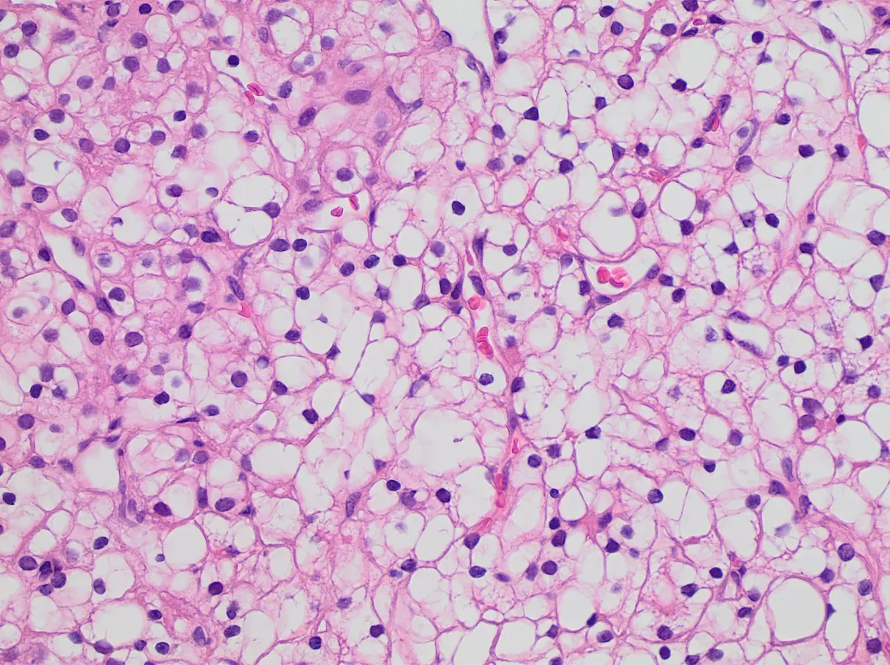
Clear cell renal cell carcinoma under the microscope
A stained tissue sample of clear cell renal cell carcinoma, grade 1, seen at intermediate magnification, with small dark nuclei in pale cells.
Open the original (2,048 × 1,532 px) (opens in a new tab)
- By
- Mikael Häggström , M.D. Author info - Reusing images - Conflicts of interest: None Mikael Häggström , M.D. Consent note : Consent from the patient or patient's relatives is regarded as redundant, because of absence of identifiable features (List of HIPAA identifiers) in the media and case information (See also HIPAA case reports guidance).
- Licence
- CC0 (opens in a new tab): free for any use, including commercial
- Source
- Wikimedia Commons (opens in a new tab)
- Tags
- cancer cells, histopathology, microscope, tissue
Credit (appreciated, not required)
Photo: Mikael Häggström , M.D. Author info - Reusing images - Conflicts of interest: None Mikael Häggström , M.D. Consent note : Consent from the patient or patient's relatives is regarded as redundant, because of absence of identifiable features (List of HIPAA identifiers) in the media and case information (See also HIPAA case reports guidance). via Pixy (https://pixy.org/673154/)